In October 2023, with the support of the National Science Foundation (NSF) and the U.S. Department of Energy’s Office of Science, researchers from Cornell University conducted an experiment to explore how microstructure emerges in a 3D-printed metal alloy by using multiple X-rays on the material while it was being printed, supported by the National Science Foundation (NSF) and the U.S. Department of Energy’s Office of Science.
The purpose was to understand how localized microscale phenomena such as bending, fragmentation, and oscillation were created in real-time using thermomechanical deformation and then be able to produce their own materials consisting of such superior characteristics.
The group’s paper was published on October 10, with doctoral student Adrita Dass being the lead author.
Atieh Moridi, the paper’s senior author, stated that they always look at the microstructures after processing, but lots of information would be missed if only postmortem characterizations were conducted. However, with the help of the current tools, they could now observe those microstructural evolutions, as well as how the tiny patterns or microstructures, which dictated everything about the performance of printed parts, were formed.
The group would apply a particular powder, the nickel-based superalloy IN625, a common aerospace industry material, into the experiment.
However, to access high-energy X-rays, they brought a portable twin of their 3D-printing setup to CHEXS@CHESS, whose staff would then work with them to develop safety protocols for operating high-power lasers. The material’s structural history would then be revealed by X-ray diffraction patterns obtained during the printing process.
At the FAST beamline, a focused X-ray beam passed through the N625 as it was heated, melted, and cooled, leading to the capture of the patterns of diffraction that took place when the X-rays and the material interacted with the other.
The team then carefully analyzed raw detector images instead of focusing entirely on diffraction data as they would typically do. This rewarded them with a clearer picture of IN625’s formation and helped them identify microstructural features such as torsion, bending, and oscillation. After that, one researcher, specifically Dass, revealed that their method could also be applied to other 3D-printed metals to discover potential enhancement. For example, pulsing a laser beam would increase fragmentation, reduce the size of its grain, and strengthen the material.
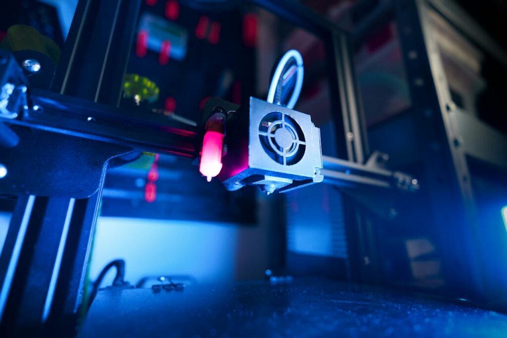
Photo by Jakub Zerdzicki on Pexels
Article Source: Cornell University